
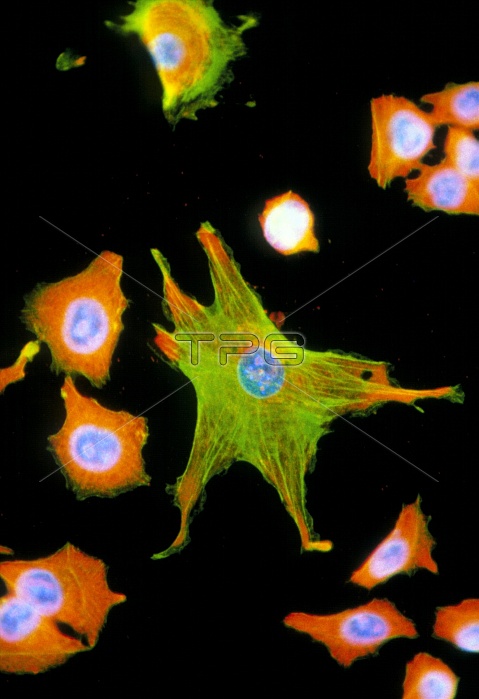
Immunofluorescent Light Micrograph of melanoma cancer cells and fibroblasts, cultured from a human tumour. A fibroblast (star-shaped cell, at centre), forms connective tissue to support other cells. Actin fibres (green) around it help provide this supportive network. Smaller melanoma cells have a large nucleus (blue); cytoplasm is orange. Derived from melanin-forming cells of the skin, they are highly malignant & divide rapidly (seen at upper right). Immunofluorescence is a staining technique which uses antibodies to attach fluores- cent dyes to specific tissues and to molecules within the cell.Mag:x200 at35mm,x340 at 6x4.5size.
| px | px | dpi | = | cm | x | cm | = | MB |
Details
Creative#:
TOP10197077
Source:
達志影像
Authorization Type:
RM
Release Information:
須由TPG 完整授權
Model Release:
N/A
Property Release:
N/A
Right to Privacy:
No
Same folder images:
 Loading