
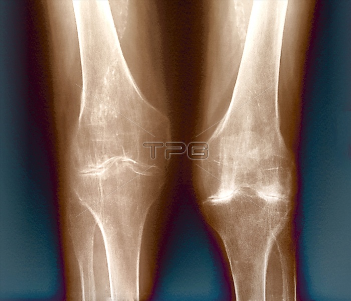
Arthritic knees. Coloured X-ray of the knees of a 59 year old with osteoarthritis. The femurs (thigh bones) are seen at top, they join to the tibias (shin bones) at bottom. The thin bones behind the tibias are the fibulas. Osteoarthritis is a joint disease caused by mechanical wear and tear. In a normal knee joint, a clear space should be seen between the femur and tibia. Osteoarthritis has led to the loss of the smooth joint surface, and of the separating cartilage. Deposits of calcium crystals (bright white) have formed in the cartilage, a common complication. The only treatment is with painkillers, or surgery in extreme cases.
| px | px | dpi | = | cm | x | cm | = | MB |
Details
Creative#:
TOP10195792
Source:
達志影像
Authorization Type:
RM
Release Information:
須由TPG 完整授權
Model Release:
N/A
Property Release:
N/A
Right to Privacy:
No
Same folder images:
 Loading