
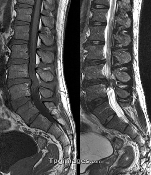
Spinal nerve pain. Sagittal magnetic resonance imaging (MRI) scans of the lumbosacral spine in a 40-year-old patient with spinal nerve pain (sciatica). The image at left is T1-weighted, with the image at right being T2-weighted. The scans pass vertically through the spinal cord and backbone (mostly to left of spinal cord), with the front of the spine at left. The pain is caused by pressure on the spinal nerves at the L4-L5 vertebral juncture, causing the bulge seen just below centre on each scan. Causes of this condition include herniated intervertebral discs.
| px | px | dpi | = | cm | x | cm | = | MB |
Details
Creative#:
TOP06670723
Source:
達志影像
Authorization Type:
RM
Release Information:
須由TPG 完整授權
Model Release:
NO
Property Release:
NO
Right to Privacy:
No
Same folder images:
sciaticadisorderconditionnervehumanbodyspinalcordspinebackbonebackjointmedicineneurologyarthrologypatientadult4040smriscanscannermonochromeblack-and-white2twoabnormalunhealthydiagnosisdiagnosticdiagnosticsmagneticresonanceimagingmedicalneuralbonebonesvertebravertebraeintervertebraldiscdiscsslippeddischerniateddiscneurologicalpainpainfultrappednervefortieslumbarlowerbackspinallateralsideviewprofilesagittalpairduolumbosacrall4l5t1-weightedt1weightedt2-weightedt2weighted"
"24040sabnormaladultarthrologybackbackbackboneblack-and-whitebodybonebonesconditioncorddiagnosisdiagnosticdiagnosticsdiscdiscdiscdiscsdisorderduofortiesherniatedhumanimagingintervertebraljointl4l5laterallowerlumbarlumbosacralmagneticmedicalmedicinemonochromemrinervenerveneuralneurologicalneurologypainpainfulpairpatientprofileresonancesagittalscanscannersciaticasideslippedspinalspinalspinet1t1-weightedt2t2-weightedtrappedtwounhealthyvertebravertebraeviewweightedweighted
 Loading