
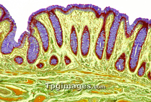
Colon lining. Coloured light micrograph of a section through a human colon. The lumen (white) of the colon is at top and the smooth muscle layer facilitating the passage of faeces is at bottom (orange and green). The colon is lined with intestinal villi containing crypts of Lieberkuhn, elongated pockets that lie in the vascular submucosa (pale green). These glands contain two types of cells (both purple): goblet cells, which secrete a lubricant into the colon which aids the movement of faeces; and absorptive columnar epithelial cells, which absorb water from the faeces. The nuclei of these cells are dark red. The number and shape of the glands greatly increases the surface area for absorption. Magnification: x300 when printed 10cm wide.
| px | px | dpi | = | cm | x | cm | = | MB |
Details
Creative#:
TOP03221903
Source:
達志影像
Authorization Type:
RM
Release Information:
須由TPG 完整授權
Model Release:
N/A
Property Release:
N/A
Right to Privacy:
No
Same folder images:
 Loading