
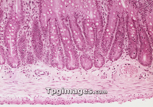
Small intestine wall. Light micrograph of a section through the wall of the human small intestine. Making up the upper half of the frame are the crypts of Lieberkuhn (long, tubular) which lie at the base of the intestinal villi (unseen). They contain numerous mucus-producing goblet cells (white, round). Just below and between the crypts is the vascular submucosa. Beneath is the circular muscle layer lying above the longitudinal muscle layer. These layers of smooth muscle are responsible for the peristaltic action of the intestine. The thin outermost layer (at bottom) is the serosa composed of connective tissue. Magnification: x200 at 35mm size.
| px | px | dpi | = | cm | x | cm | = | MB |
Details
Creative#:
TOP03221754
Source:
達志影像
Authorization Type:
RM
Release Information:
須由TPG 完整授權
Model Release:
N/A
Property Release:
N/A
Right to Privacy:
No
Same folder images:
 Loading