
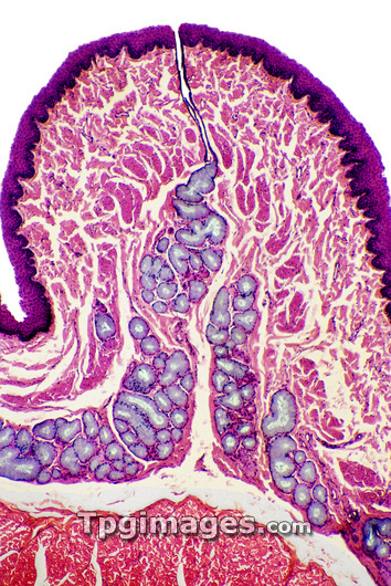
Oesophagus wall. Coloured light micrograph of a section through the human oesophagus, which passes food from the mouth to the stomach. The oesophagus lumen (white, top) is lined by a layer of stratified squamous epithelium (dark purple). This protects underlying tissue and enables the passage of solid material by the lubricant action of its glandular secretion. Next is a supporting layer of connective tissue (pink), the lamina propria , which is rich in elastin fibres. A mucus duct (upper centre) can be seen running down these layers to join the submucosa, which contains mucous glands (purple) for lubricating the oesophagus as food passes through it. At bottom is a thick layer of circular and longitudinal muscles (red). Magnification: x80 when printed 10cm tall.
| px | px | dpi | = | cm | x | cm | = | MB |
Details
Creative#:
TOP03221741
Source:
達志影像
Authorization Type:
RM
Release Information:
須由TPG 完整授權
Model Release:
N/A
Property Release:
N/A
Right to Privacy:
No
Same folder images:
 Loading