
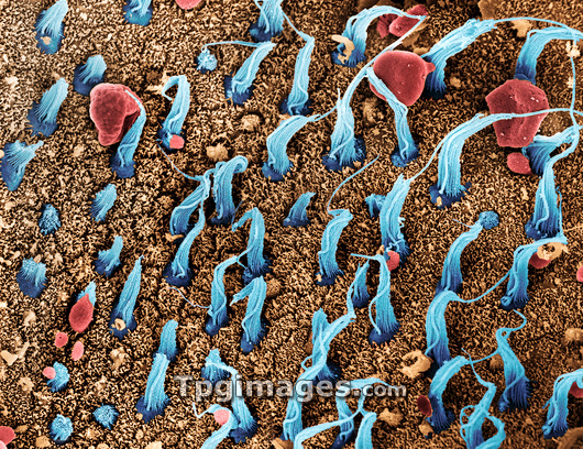
Inner ear sensory cells. Coloured scanning electron micrograph (SEM) of bundles of cilia (hair cells, blue) situated in the macula utriculi within the human inner ear. Each bundle comprises many stereocilia and one long kinocilium. They detect directional movement, such as the tilting of the head. The cilia are immersed in a fluid, called the endolymph. Currents in this fluid displace the position of the cilia, resulting in the production of electrical pulses, which are sent to the brain along the vestibular nerve. The surrounding support cells are covered in projections called microvilli (brown). Otoliths (red) are also seen. These crystals also help sense balance.
| px | px | dpi | = | cm | x | cm | = | MB |
Details
Creative#:
TOP03221633
Source:
達志影像
Authorization Type:
RM
Release Information:
須由TPG 完整授權
Model Release:
N/A
Property Release:
N/A
Right to Privacy:
No
Same folder images:
 Loading