
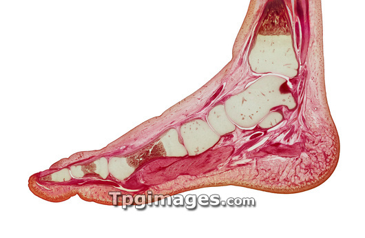
Foetal foot. Macro photograph of a sagittal section through the foot of a 7 month old foetus at the level of the big toe. At top right is the tibia (shin bone), which articulates with the talus to form part of the ankle joint. Bottom right of the talus is the calcaneus (heel bone). To the left of the talus is the navicular bone, with the medial cuneiform bone to the left of that. Collectively these bones are part of the tarsal group of bones. The metatarsal links the tarsal bones to the phalanges of the toe. The big toe consists of a proximal (right) and a distal (left) phalanx. Bone growth plates (brown) are seen in the phalanges and metatarsals. This slide dates from 1897.
| px | px | dpi | = | cm | x | cm | = | MB |
Details
Creative#:
TOP03220826
Source:
達志影像
Authorization Type:
RM
Release Information:
須由TPG 完整授權
Model Release:
N/A
Property Release:
N/A
Right to Privacy:
No
Same folder images:
 Loading