
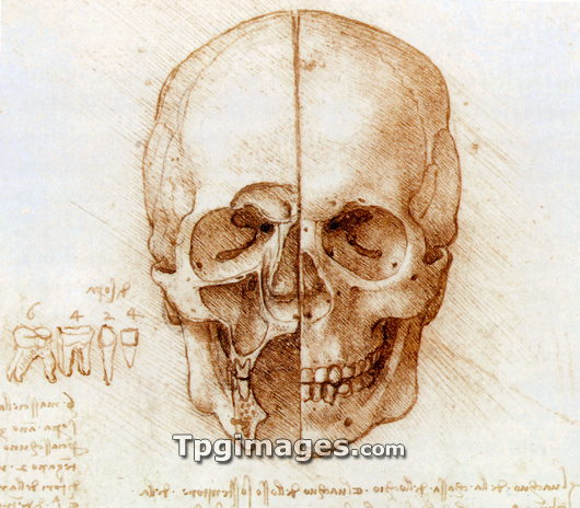
Skull anatomy by Leonardo da Vinci. Historical artwork and notes on the anatomy of the human skull and teeth, by the Italian artist and scientist Leonardo da Vinci (1452-1519). This bisected skull shows the external structure (right), and dissected facial sinuses (left), the air-filled spaces inside the bones of the face. The diagram at lower left shows the teeth present in one half of the mouth: 4 incisors, 2 canines, 4 pre-molars, and 6 molars. Da Vinci was the first anatomist known to have correctly noted the number and root structure of human teeth. The notes are an example of his mirror writing, which was written backwards from right to left, and could be read in a mirror.
| px | px | dpi | = | cm | x | cm | = | MB |
Details
Creative#:
TOP03220449
Source:
達志影像
Authorization Type:
RM
Release Information:
須由TPG 完整授權
Model Release:
N/A
Property Release:
N/A
Right to Privacy:
No
Same folder images:
 Loading