
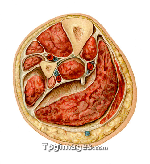
Transverse section of lower leg. Historical anatomical artwork of a transverse section through the middle third of a lower leg, showing the internal structures surrounded by layers of fat (yellow) and skin (pink). The two bones are the tibia (shin bone, top) and the fibula (centre left). Several sets of muscles (red) are shown, surrounded by muscle membranes (white), including the large calf muscle (lower right). Nerves (white), arteries (red) and veins (blue) are also shown. The main nerve (centre) is the posterior tibial nerve, with the posterior tibial veins and artery to its right. Above the fibula are the anterior tibial artery and vein, with the deep peroneal nerve. Below the fibula are the peroneal artery and vein. Artwork from Atlas and Epitome of Operative Surgery (1898, Otto Zuckerkandl).
| px | px | dpi | = | cm | x | cm | = | MB |
Details
Creative#:
TOP03220415
Source:
達志影像
Authorization Type:
RM
Release Information:
須由TPG 完整授權
Model Release:
N/A
Property Release:
N/A
Right to Privacy:
No
Same folder images:
 Loading